Molecular Fingerprints Converge into a Health Map
©2025 Hangzhou Well-healthcare Technologies Co., Ltd Copyright.


MS Detection Chip
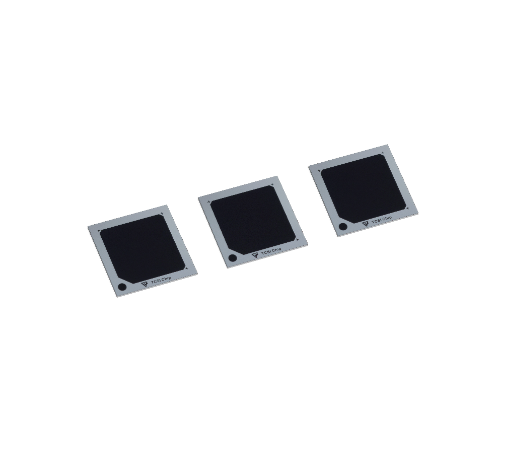
TCSI Array® Imaging MS Chip
TCSI Array® Imaging MS Chip is the first disposable silicon-based target plate in China designed for MALDI imaging studies. It enables simultaneous molecular weight calibration and imaging analysis. Suitable for analyzing tissue samples obtained through biopsy, microscopy, and intraoperative procedures, as well as fingerprints, plant sections, and more.

Product Consultation
Matrix-free detection, simplifying the experimental workflow
High ionization efficiency with excellent fidelity
Integrated molecular weight calibration sites for high mass accuracy
Compatible with various imaging platforms, customizable and adaptable for further development based on user needs
-Tissue Imaging & Endogenous Spatial Metabolomics/Lipidomics Analysis
-Fingerprint Imaging & Explosive Residue Detection
-Drug Distribution Analysis
Compatible with vacuum-based mass spectrometry imaging and customizable in size and parameters to fit different instruments.
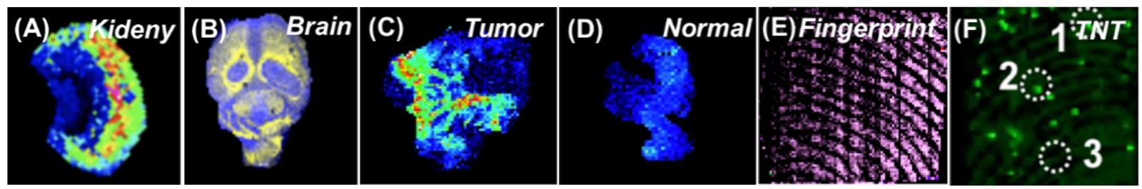
A. Mouse Kidney Tissue Imaging B. Mouse Brain Tissue Imaging C/D. Lung Cancer Tissue Imaging (C) and Adjacent Normal Tissue Imaging (D)
E. Latent Fingerprint Imaging F. Explosive Residue Imaging in Fingerprints
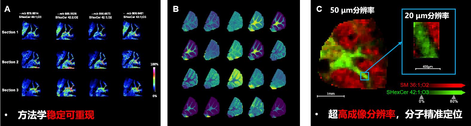
Cerebellum Tissue Imaging Results on the TCSI Array® Mass Spectrometry Chip:
(A) Imaging results of parallel cerebellum tissue sections.
(B) Imaging distribution of different molecules in the cerebellum region.
(C) Molecular imaging in the cerebellum region at 50 μm and 20 μm resolution.
Covers a wide range of metabolic molecules
Imaging Resolution
Phospholipids: PI, PE, PG, PS, PC Sphingolipids: SM, SHexCer, GalCer Neutral lipids: ST, TG, DG
20-200 μm
| Product | Compatible Mass Spectrometers | Application | Specifications | Model | Item NO. |
|---|---|---|---|---|---|
TCSI Array® Imaging MS Chip | autoflex max、ultraflex maX、 rapidflex maX、MALDI2 | Used for mass spectrometry imaging and analysis of plant and animal tissues, with specifications selected based on the area of the sample to be analyzed | 15×15 mm | TCSIArray02 | HR5006 |